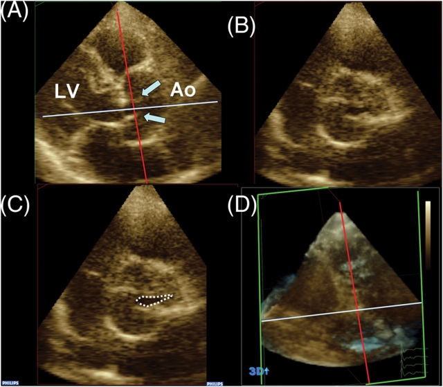
Figure 3.
Real-time three-dimensional echocardiographic aortic valve area (AVA) by planimetry. Cropping allows alignment of short-axis plane (B and C) at the narrowest orifice as visualized in long axis plane (A). Arrows point to the aortic valve leaflets in systole. Cropping planes are shown in 3D set for reference (D). LV, left ventricle; Ao, Ascending Aorta.

