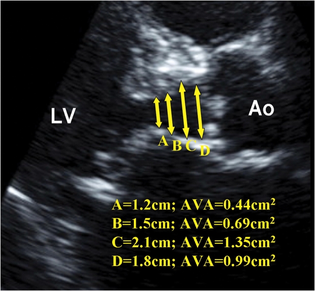
Figure 6.
Two-dimensional echocardiography at the parasternal long-axis view illustrating how upper septal hypertrophy distorting the left ventricular outflow tract geometry might lead to erroneous and wide variation of aortic valve area (AVA) calculation by two-dimensional continuity equation. LV, left ventricle; Ao, Aorta.

