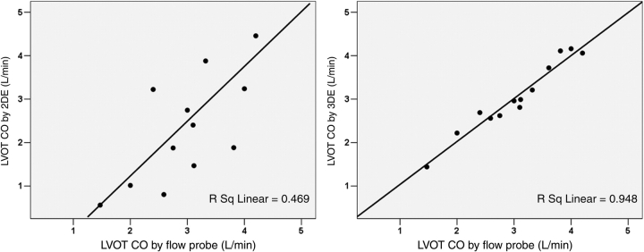
Figure 7.
Correlations between the left ventricular outflow tract (LVOT) cardiac output (CO) derived from two-dimensional echocardiography (2DE) Doppler (left panel) and three-dimensional echocardiography (3DE) colour Doppler (right panel) vs. the referenced flow probe measurements. These were obtained from sheep models with distorted LVOT geometry.

