
Figure 1:
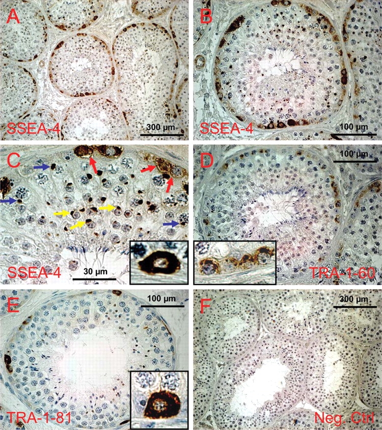
Expression of glycan stem-cell markers in the testis of C. jacchus as revealed by immunohistochemistry.
(a) SSEA-4 expression, overview. (b) SSEA-4 expression in a roundish cross-section of a seminiferous tubule. All labeled cells are in contact with the basal lamina. (c) Higher magnification of the upper part of the tubule shown in (b). Spermatocytes show a strongly stained perinuclear dot (blue arrows) which most likely represents the XY body. In spermatids the acrosome is stained (yellow arrows). Spermatogonia exhibit homogenous cytoplasmic and membrane staining (red arrows and inset), while the nuclei are devoid of stain. (d) A tubule showing TRA-1-60 positive spermatogonia, which are also invariably in contact with the basal membrane. However, subcellular distribution of the stain is clearly different from that of the other glycan markers. TRA-1-60 antigen is concentrated in the apical part of the spermatogonia (inset). Decreased stain was also detected in the lateral parts of the cells, while no stain was found in the cytoplasmic and membrane compartment between the nucleus and the basal membrane (inset). (e) Expression of TRA-1-81. Fewer cells are positive compared to the other glycan markers and those cells labeled are almost exclusively single cells. Subcellular distribution of the stain resembles SSEA-4, i.e. the cytoplasm is homogenously stained, but the nucleus is free of stain (inset). (f) Negative control for all antibodies (all from mouse) used.