Fig. 1.
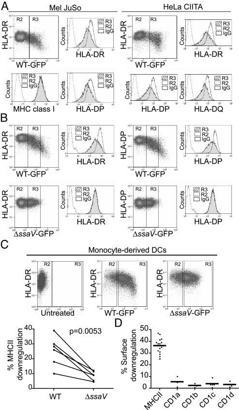
Salmonella infection results in selective loss of surface MHC class II expression. (A) Mel JuSo and HeLa-CIITA cells were infected with Salmonella-GFP and HLA-DP, -DQ, and -DR expression was examined in GFP positive (infected, R3) and GFP negative (uninfected, R2) cells. Panels show representative FACS dot plots of HLA-DR against GFP expression, in infected Mel JuSo and HeLa-CIITA cells. Histograms show surface DR, DP, DQ, and MHC class I expression in infected (hatched) and uninfected (clear) cells. Isotype control is represented by a dashed line. Data are representative of more than 3 independent experiments. (B) Mel JuSo were infected with WT and ssaV Salmonella-GFP strains and DP and DR examined in infected and uninfected cells. Panels show representative FACS dot plots of HLA-DR and HLA-DP expression in cells infected with similar numbers of bacteria (R3). Histograms show surface DR and DP expression in infected (hatched) and uninfected (clear) cells. Isotype control is represented by the dashed line. Data are representative of more than 3 independent experiments. (C) Immature DCs were left uninfected or infected with WT or ssaV strains for 30 min and the entire population of cells processed for FACS analysis 16–20 h post infection. DR is only reduced by infection with WT Salmonella (upper panels). Comparison of DR down-regulation in DCs derived from 6 independent donors infected with WT or ssaV strains is shown in the lower panel. (D). Mel JuSo cells, stably expressing either CD1a, CD1b, CD1c, or CD1d, were infected with WT Salmonella. Surface expression of each CD1 isotype, together with HLA-DR, was measured by FACS. Dots show the percentage surface down-regulation of both HLA-DR and CD1. Error bars, means.

