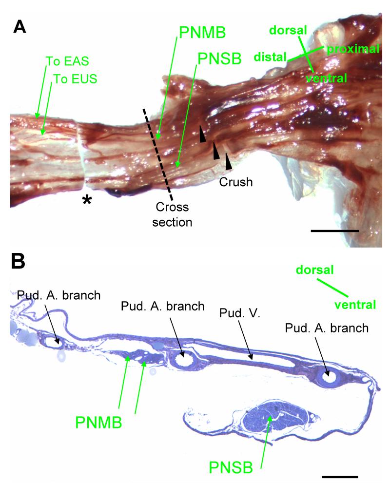
Fig. 4.
Nerves in Alcock’s canal in female rats. A. the pudendal nerve showing the location of nerve crush. The sample for embedding was indicated between the dashed line and * (the distal cut section). EUS: external urinary sphincter; EAS: external anal sphincter; PNMB: pudendal nerve motor branch; PNSB: pudendal nerve sensory branch. Bar = 1 mm. B. Alcock’s canal cross section at the proximal end of the sample shown in A (the dashed line), showing the PNMB, the PNSB, the pudendal artery (Pud. A), and the pudendal vein (Pud. V). Bar = 250 μm.

