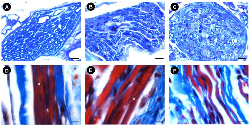
Fig. 5.
Examples of the pudendal nerve motor branch (PNMB) and external urethral sphincter (EUS) in controls (A, D); 6 weeks after PNC+VD (B, E), and 6 weeks after PNT (C, F). Nerve stain (A, B, C) = toluidine blue; Muscle stain (D, E, F) = Masson’s trichrome. * indicates the cross striation (D, E). Bar = 5 μm.

