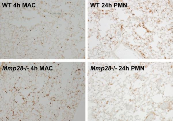
Fig. 6. Lung histology after P. aeruginosa inhalation.

These are representative images demonstrating enhanced macrophage influx into the lung of Mmp28−/− mice at 4 h post-infection with P. aeruginosa (Bottom Left), and reduced neutrophil influx into the lung of Mmp28−/− mice 24 h post-infection with P. aeruginosa (Bottom Right) compared to WT controls (Top). Lungs are fixed in 10% formalin, paraffin-embedded and stained with anti-mouse MAC-2 antibody (left) or anti-neutrophil antibody (right). Secondary antibody was HRP-conjugated donkey anti-rat. Secondary antibodies were labeled with the Vectastain ABC kit and colorimetric detection was done with diaminobenzidine staining.
