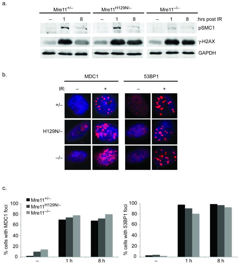
Figure 4. Independence of γ-H2AX-MDC1-53BP1 and MRN.
(a) Distinct requirements for phosphorylation of SMC1 versus H2AX during the response to DSBs. Western blot analyses of the indicated proteins (right) prior to 10 Gy IR (−), and 1 or 8 hours post-IR. Phosphorylated SMC1 is used as a control to demonstrate functional MRN deficiency9. GAPDH is a protein loading control. (b) Representative immunoflourescent foci prior to (−), or 8 hours after (+) 10 Gy IR. (c) The formation of MDC1 and 53BP1 foci at 1 and 8 h post-IR does not require Mre11 nuclease activities or MRN. Bar graphs represent quantification of MDC1 and 53BP1 foci-positive cells at the indicated times (X axis) after 10 Gy IR. (−) no IR. Positive cells defined as > 5 foci.

