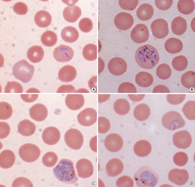
Fig. 1.
Wright-Giemsa-stained peripheral blood smear (magnification, ×1,000). (A) a ring form in an enlarged red blood cell with fimbriated margin, (B) a schizont with eight merozoites distributed like a daisy-head, (C) a microgametocyte with eccentric and dispersed chromatin, (D) a macrogametocyte with eccentric and condensed chromatin.

