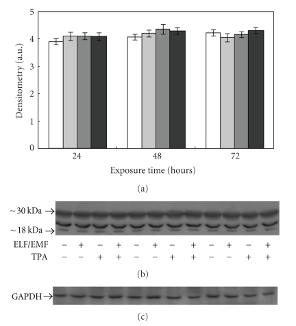
Figure 7.
Effect of ELF-EMFs exposure and TPA treatment on 20S proteasome levels. Representative autoradiography of 20S proteasome in Caco cells exposed to ELF-EMFs. The densitometric analysis from six separate blots provided for quantitative analysis of the amount of 20S “core” is presented (a) and a representative Western blot is shown in (b). Equal protein loading was verified by using an anti-GAPDH antibody (c). The immunostaining was performed using an anti-20S proteasome antibody, and the detection was executed by the Enhanced Chemiluminescence Western blot analysis system.

