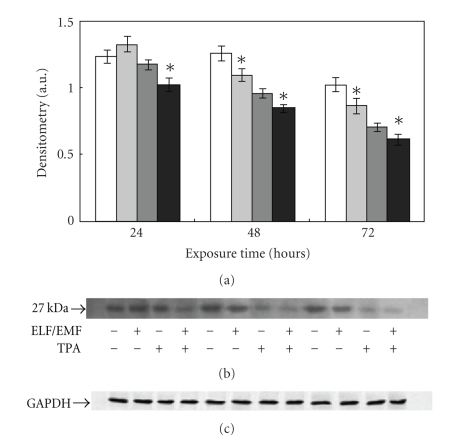
Figure 8.
Effect of ELF-EMFs exposure and TPA treatment on p27 levels. Autoradiographs of p27 expression in Caco cells exposed to ELM-EMFs. The densitometric analysis from six separate blots provided for quantitative analysis is presented (a) and a representative Western blot is shown (b). Equal protein loading was verified by using an antibody directed against GAPDH (c). Caco cells were cultured with or without 0.1 μM TPA for 24, 48, and 72 hours. After harvesting and lysing the cells, samples were subjected to SDS-PAGE and electroblotted on a polyvinylidene fluoride membrane. The immunostaining was performed using anti-p27 antibodies, and the detection was executed by the Enhanced Chemiluminescence western blotting analysis system. Data points marked with an asterisk are statistically significant compared to their respective not exposed control cells (P < .05).

