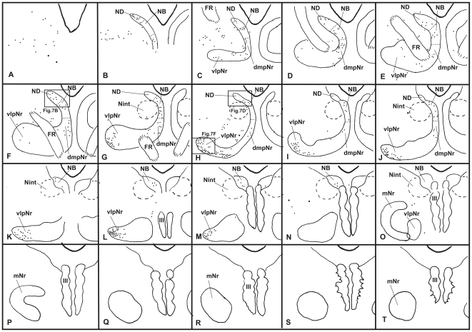
Figure 6. Distribution of WGA-HRP-labelled cells in mesodiencephalic structures (case 4).
Drawings are arranged rostrocaudally; section A is the most rostral. One dot represents one labelled cell. Large dot shows a large labelled cell. dmpNr–dorsomedial part of parvicellular red nucleus, FR–fasciculus retroflexus, mNr–magnocellular red nucleus, NB–nucleus accessorius medialis of Bechterew, ND–nucleus of Darkschewitsch, Nint–interstitial nucleus of Cajal, vlpNr–ventorlateral part of parvicellular red nucleus, III–oculomotor nucleus.

