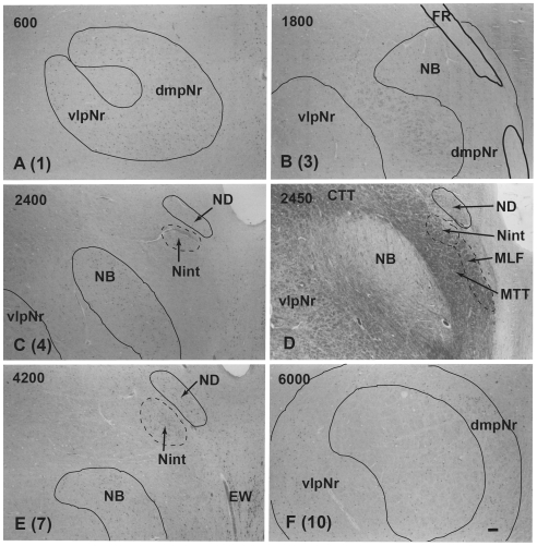
Figure 11. Human parvicellular red nucleus.
Photomicrographs showing the distribution of Nissl-stained pNr cells in A(1), B(3), C(4), E(7), and F(10) which correspond to sections 1, 3, 4, 7 and 10 of Fig. 10 and the distribution of myelinated fiber bundles stained by myelin stain in D which corresponds to section Fig. 10B. The number in the left corner of each photomicrograph is the rostrocaudal distance (in micrometers) from the rostral tip of the red nucleus. CTT–central tegmental tract, dmpNr–dorsomedial part of parvicellular red nucleus, EW–Edinger-Westphal nucleus, FR–fasciculus retroflexus, MLF–medial longitudinal fasciculus, MTT–medial tegmental tract, NB–nucleus accessorius medialis of Bechterew, ND–nucleus of Darkschewitsch, Nint–interstitial nucleus of Cajal, vlpNr–ventorlateral part of parvicellular red nucleus. Scale bar = 200 µm in F (also applies to A–E).

