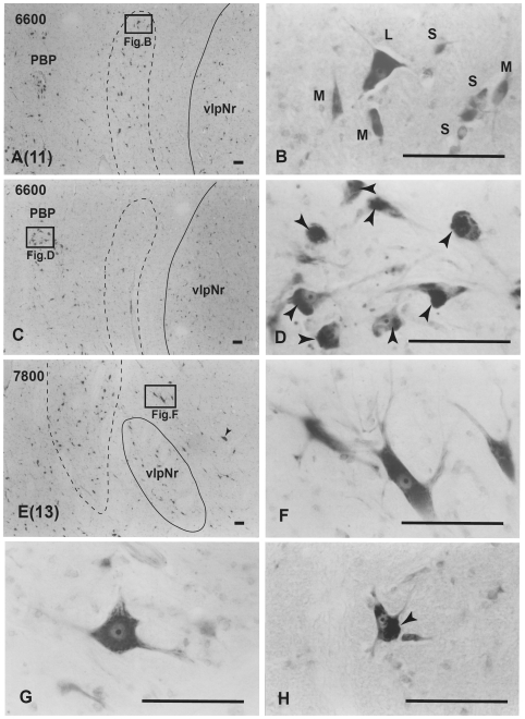
Figure 12. Human magnocellular red nucleus.
Photomicrographs showing the distribution of Nissl-stained mNr cells. A (11) and E (13) correspond to sections 11 and 13 in Fig. 10. C is a reversed photomicrograph of the contralateral side of A for comparison with C. The area indicated by a broken line is the outer layer of the semi-lunar shell of mNr (A, C and E). It exits between the capsule of the superior cerebellar peduncle and the parabrachial pigmented nucleus (PBP). B. High magnification views of large (L), medium (M) and small (S) neurons of the outer shell of mNr. D. Many pigmented cells exist in the parabrachial pigmented nucleus. Arrowheads indicate accumulation of pigment. E. Caudal end of vlpNr. Arrowhead indicates one giant neuron. F and G. Giant neurons among fibers of the superior cerebellar pedunculus. H. Large neuron contains pigments. Arrowhead indicates accumulation of pigment. The number in the left corner of each photomicrograph is the rostrocaudal distance (in micrometers) from the rostral tip of the red nucleus. PBP–parabrachial pigmented nucleus, vlpNr–ventorlateral part of parvicellular red nucleus. Scale bars = 100 µm in A–H.

