
Figure 1.
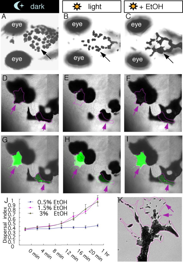
Ethanol modulates the camouflage response in larval zebrafish by stimulating melanosome dispersal. A–C, Nomarski images of 7 dpf larval zebrafish exposed to dark (A), light (B), or ethanol in light (C), showing the appearance of melanocytes on the head region. D–I, In vivo time-lapse Nomarski images (D–F) or Nomarski superimposed with fluorescence images (G–I) showing two GFP-positive melanocytes (arrows) undergoing dispersal in dark (D), aggregation in light (E), and dispersal in ethanol and light (F). J, Quantification of melanocyte dispersal upon treatment with different concentrations of ethanol. K, A snapshot from a high-magnification movie (supplemental Movie S1, available at www.jneurosci.org as supplemental material) of a cultured melanocyte, showing individual organelles inside the melanocytes, called melanosomes (arrows), undergoing aggregation.