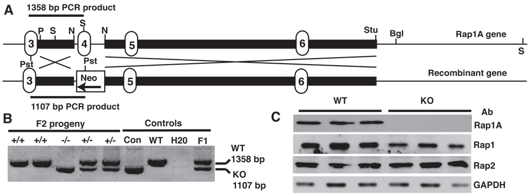
FIGURE 1. Rap1a deletion strategy and detection.
A. Knockout strategy showing replacement of exon 4 with neo resistance gene. Positions of genotyping primers are shown as well restriction enzymes used in gene manipulation or analysis. Bgl, BglII; N, NdeI; Pst, PstI; P, PvuII; S, SpeI; Stu, StuI. Numbered regions indicate location of exons, solid black bars show short and long arms used in homologous recombination.
B. PCR analysis of Rap1a null mice. Using a common primer 5’ of the targeted sequence (in exon 3) and 3’ primers complementary to either exon 4 or the neoR gene, PCR reactions detected Rap1a +/+ (upper band only) −/− (lower band only) or +/− (both bands) mice. Con, control plasmid template; F1, chimeric founder mouse.
C. Western blot of Rap expression in mouse neutrophils. Cells were blotted for Rap1a, total Rap1 or Rap2 expression using specific antibodies. Glyceraldehyde 3 phopshate dehydrogenase (GAPDH) was used as an internal loading control. Data are shown for 3 separate Rap1a +/+ and −/− mice.

