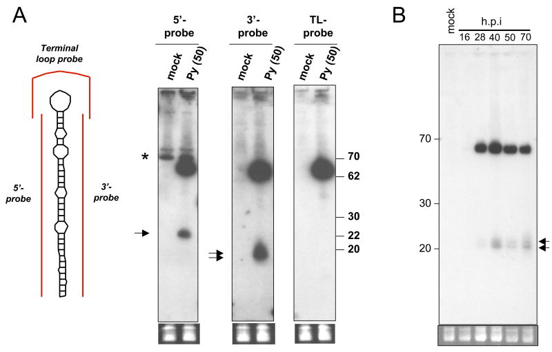
Figure 2. Northern blot confirmation of MR1 candidate.
(A) Northern blot analysis was conducted using the probes diagramed in red (left side of Fig.) on RNA harvested from mock or PyV-infected BMK cells at 50 h.p.i.. Arrows indicate bands corresponding to miRNAs. Note predominant band detected by all three probes migrating at ~65 nucleotides corresponds to the pre-miRNA. Bottom panel shows pre-transferred, low molecular weight RNAs stained with ethidium bromide as a loading control (B) Time course of pre-miRNA/miRNA accumulation (hybridized with 5′ probe).

