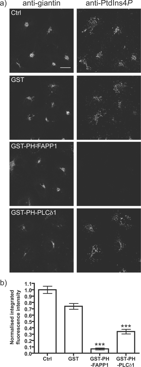
Figure 5. Competition for Golgi PtdIns4P staining by PH-FAPP1.
(a) The indicated GST-tagged fusion proteins (10 μM) were included with the primary anti-PtdIns4P antibody and anti-giantin antibodies applied to COS-7 cells. Staining conditions are as Figure 3. Single confocal optical sections are shown. Scale bar=20 μm. (b) Quantification of anti-PtdIns4P antibody labelling (n=207). See the Experimental section for details. ***P<0.001.

