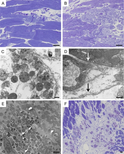
Figure 8.
Ultrastructural features of cardiomyocyte necrosis in Tg-NS/H mice. (A) Focal contraction band necrosis on a semithin section. Bar, 10 µm. (B) Myocytolysis on a semithin section. Bar, 10 µm. (C) Disgregation of cytoplasm with loss of myofilaments and woolly bodies inside swollen mitochondria (m), features in keeping with necrotic cell death. Bar, 500 nm. (D) Loss of sarcolemma integrity in a cardiomyocyte (black arrow) with cytoplasmic disgregation, close to abnormal cardiomyocyte with intact sarcolemma (white arrow). Bar, 500 nm. (E) Mineralization of mitochondria. Bar, 2 µm. (F) Spotty calcification within fibrous tissue on a semithin section. Bar, 10 µm.

