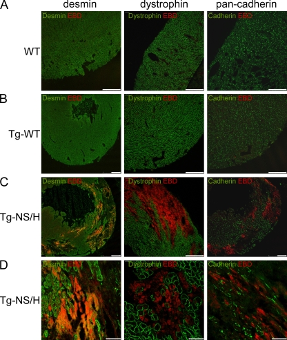
Figure 9.
Immunohistochemical analysis of cardiac cryosections from 2–3-wk-old mice injected with EBD. (A and B) WT and Tg-WT hearts show normal distribution of desmin, dystrophin, and pan-cadherin in the ventricles, and no EBD-positive cells. Bars, 250 µm. (C and D) In contrast, hearts from Tg-NS mice display large areas of ventricular myocardium lacking desmin, dystrophin, and pan-cadherin. These abnormal areas contained myocytes positive for EBD, indicating increased cell membrane permeability and loss of myocyte integrity and viability. Bars: (C) 250 µm; (D) 50 µm.

