Figure 6.
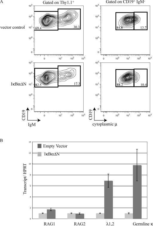
B cell development in IκBαΔN-infected IL-7 bone marrow cultures. (A) Bone marrow IL-7 cultures were infected with an IκBαΔN retrovirus or an empty vector control. 3 d after infection, cells were surface stained with Thy1.1 (a marker of viral infection), CD19, and IgM antibodies. The cells were fixed, permeabilized, stained with IgM antibody, and then analyzed by flow cytometry. The top shows the empty vector control infection and the bottom shows IκBαΔN-infected cells. The left displays CD19 versus IgM gated on Thy1.1+ cells. The right displays CD19 verses cytoplasmic μ gated on CD19+IgM−. (B) Real-time RT-PCR analysis of the indicated transcripts using RNA purified from Thy1.1-sorted IκBαΔN or empty vector retrovirally transduced pro–/pre–B cells. All transcripts were normalized to HPRT transcription and data from each IκBαΔN sample was arbitrarily set to 1, with error bars indicating range of triplicate assays. These experiments were repeated twice using bone marrow pooled from six mice.

