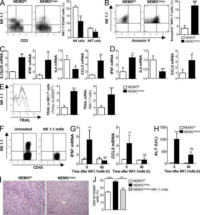
Figure 3.
Hepatocyte-specific deletion of NEMO promotes spontaneous activation of liver NK/NKT cells, and administration of NK1.1-depleting mAb attenuates the damaging phenotype in NEMOΔhepa mice. (A) FACS analysis revealed a lower number of NK1.1+CD3− and NK1.1+CD3+ cells in NEMOΔhepa mice. The graph represents the percentage of NK1.1+ cells related to the percentage of CD45+ cells in the liver (B) FACS analysis of NK1.1+/annexin V+ related to % of CD45+ cells revealed strong apoptosis of NK1.1+ cells in livers from NEMOΔhepa mice. (C) RT-PCR showed strong expression of IL-12 (p35), IFN-γ, and CCL5 but lower IL-4 mRNA in livers. Data are presented as times versus NEMOf/f untreated. (D) mRNA analysis of isolated NK1.1+ cells confirmed cell activation and increased cytokine expression. (E) FACS analysis showed stronger TRAIL expression on NK1.1+ cells from livers from NEMOΔhepa mice. mRNA analysis on isolated NK1.1+ cells confirmed this. (F and G) FACS analysis proved effective depletion of NK1.1+ cells (F) 40 h after NK1.1 mAb administration that reduced IFN-γ and CCL5 mRNA levels (G). (H–J) Serum ALT (H), H&E staining (I), and FACS analysis (J; CD11b+ cells) were used as markers of liver damage and inflammation. Bars, 50 µm. All data are representative of three independent experiments. n = 4. *, P < 0.05; **, P < 0.01; ***, P < 0.001 (NEMOf/f vs. NEMOΔhepa). §§, P < 0.01 (NEMOΔhepa vs. Nk1.1mAb/NEMOΔhepa). Error bars represent SD.

