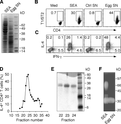
Figure 1.
S. mansoni egg culture supernatants promote bystander Th2 polarization. (A) Protein composition of SEA and egg supernatants evaluated by Coomassie blue–stained SDS-PAGE. (B and C) Naive DO.11.10 Tg CD4+ lymphocytes were cultured with DC and OVA peptide with or without 40 µg/ml SEA, 30 µg/ml egg supernatant, or control supernatants from mock cultures containing no eggs. After restimulation with PMA/ionomycin, cells were stained for CD4 and T1/ST2 (B) or CD4 and IL-4 plus IFN-γ (C), respectively. The FACS dot plots shown are gated on CD4+ T lymphocytes (percentages are shown). The experiment shown is representative of more than five performed. (D) The frequency of IL-4+ DO.11.10 T cells determined by intracellular cytokine staining (ICS) in response to gel filtration fractions of egg culture supernatants. (E) Coomassie blue–stained SDS-PAGE of fractions 22–24 from the column shown in D. Results are representative of two independent experiments performed with different egg culture supernatant preparations. (F) In situ ribonuclease activity of a 32-kD protein in both SEA and egg supernatants detected by zymogram gel.

