Figure 6.
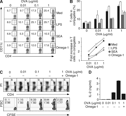
Omega-1–exposed DCs are less efficient in forming conjugates with CD4+ T cells. (A and B) BMDCs were cultured on nontreated plates with the same stimuli as in Fig. 5 C, pulsed with increasing amounts of OVA peptide, and incubated with OT-II Tg CD4+ T cells. The frequency of CD4+ T cell–forming conjugates with DC was measured by FACS. (A) A representative set of contour plots gated on CD4+ T lymphocytes (percentages are shown). (B) Quantification of T cells forming conjugates with DCs (means ± SD for duplicates) from one experiment (top). Fold increase in the frequency of OT-II Tg CD4+ T cells in conjugates with DCs was calculated as a function of peptide concentration for each type of DC based on the results from four independent experiments performed, of which two included omega-1 (bottom). (C and D) OT-II Tg T cells were cultured in the presence of DCs and increasing concentrations of OVA peptide for 36 h. Cultures with 1 µg/ml of peptide were tested with and without 0.5 µg/ml omega-1. (C) Viability of CD4+ T cells determined by PI staining of ungated populations (top). The contour plots gated on the CD4+ PI− population shows the percentage of cells that have completed the first cell cycle as determined by CFSE dilution (bottom). Cell size is indicated by forward scatter (FSC) on the y axis. (D) IL-2 in corresponding cultures measured by ELISA. Data shown are the mean concentrations ± SD. The data shown in C and D are representative of two experiments.

