Figure 7.
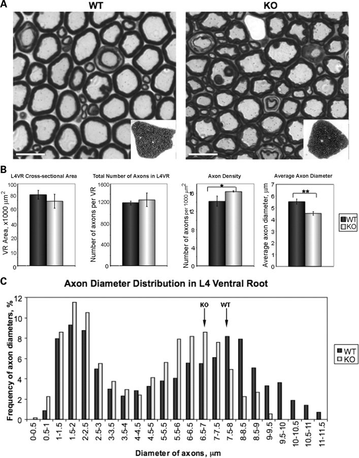
Diameters of myelinated axons are reduced in T32KO. (A) Semi-thin sections of L4 VRs from WT and KO stained with toluidine blue. L4 VRs were dissected from three WT and three KO, 8.5-month-old animals. Representative micrographs are shown. Scale bar is 10 µm. Insets show low magnification images of the sections. (B) Comparison of morphometric data from KO and WT L4 VRs. Cross-sectional area of L4 VRs is represented as the mean for each genotype ± SEM. Total number of axons in L4 VRs was calculated using VR cross-sectional area and number of axons per 1000 µm2. Axon density was calculated as the number of axons per 1000 µm2. T32KO axonal density is increased by 15%, compared with WT (*P < 0.05). Average axon diameter of T32KO is reduces by 22%, compared with WT (**P < 0.0001). (C) Histogram shows axon diameter distribution in L4 VR. Axon diameters were measured in three random areas of VR containing a total of 200–240 axons. The number of axons with certain diameter was expressed as the percent of total number of axons measured in each sample (three KO and three WT). Note that the peak corresponding to large diameter axons is shifted to the left in T32KO, compared with WT (arrows). Axons with diameter more than 9.5 µm were not observed in T32KO L4 VR.

