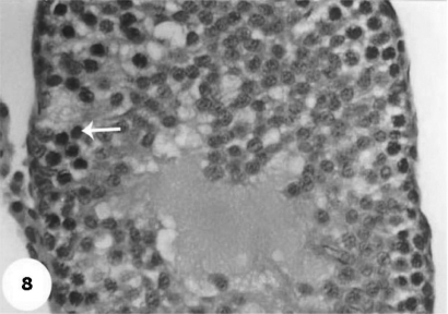
Figure 8.
A magnified photomicrograph for section of testis of AFB1-injected group shows absence of spermatids in lumen and a marked mitotic divisions and pyknotic of some spermatogonic nuclei (arrow), in addition to absence of the spermatozoa within the lumen after 2 weeks of injection. H & E × 10.

