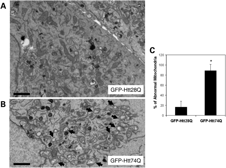
Figure 2.
Electron microscopic examination of mitochondria in HeLa cells stably expressing GFP-Htt polyglutamine fusion proteins. HeLa cell lines stably expressing either GFP-Htt28Q or GFP-Htt74Q proteins were treated with 25 µm H2O2 for 30 min, then fixed and examined by electron microscopy. (A) Representative electron microscopic image of a section taken through two adjacent cells in the GFP-Htt28Q-expressing cell line. Note the variation in size of the mitochondria as well as the close-packed cristae. Bar: 2 µm. (B) Representative electron microscopic image of a section taken through the GFP-Htt74Q expressing cell line. Note that the presence of many small damaged mitochondria that have highly disorganized cristae and whose mitochondrial matrix is considerably less electron-dense (indicated by arrows). Bar: 2 µm. (C) Quantification of the number of abnormal mitochondria (defined as containing both disorganized cristae and a less electron-dense matrix) by electron microscopic examination in the GFP-Htt28Q and GFP-Htt74Q expression lines. For this quantification, 529 and 809 mitochondria seen in 10 and 16 different GFP-Htt28Q- and GFP-Htt74Q cells, respectively, were counted. Data are shown as mean ± SDM (*P < 0.001).

