
Figure 4.
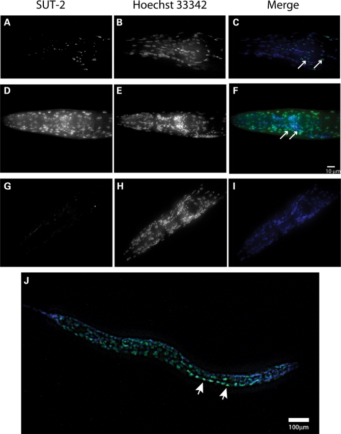
SUT-2 protein is expressed primarily in the nucleus of neurons and other cell types. (A–C) Expression of SUT-2::GFP reporter transgene in the head of an adult N2 animal. (A) SUT-2::GFP fluorescence. (B) Hoechst 33342 nuclear marker fluorescence staining. (C) Merged images of (A and B), green is GFP-fluorescence, blue is Hoechst 33342. Arrows represent nerve ring neurons. (D–F) Depict immunocytochemical staining of wild-type worms with anti-SUT-2 specific antibodies (D) and Hoechst 33342 nuclear marker (E) and a Merged image (F) (green = SUT-2, blue = Hoechst 33342). Arrows represent nerve ring neurons. (G–I) Depict immunocytochemical staining of sut-2(bk741) worms with anti-SUT-2 specific antibodies and Alexa 488 fluorescent secondary antibodies (G) and Hoechst 33342 nuclear marker (H) and a merged image (I). (B and C) Are high magnification views of the heads of adult sut-2(bk87) and sut-2(bk741) animals, respectively. (D) Shows a high magnification view of the head of an adult N2 animal. (J) Shows an L2 stage wild-type larvae stained with SUT-2 specific antibodies (green) and Hoechst 33342 (blue). Arrows represent nerve ring neurons. Arrowheads indicate normal ventral cord neurons with SUT-2 protein staining.