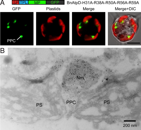
Fig. 4.
Immnocytochemical localization of a GFP fusion protein in the PPC. (A) Confocal images of a L. amoebiformis cell transformed with pBnAtpD-H31-R38A-R50A-R56A-R59A+GFP, showing the GFP localization in the PPC. (Scale bar, 5 μm.) (B) An immunoelectron micrograph of a plastid in a cell transformed with the pBnAtpD-H31A-R38A-R50A-R56A-R59A construct, showing the accumulation of conjugated gold particles (10 nm) in the PPC including a nucleomorph. Nm, nucleomorph.

