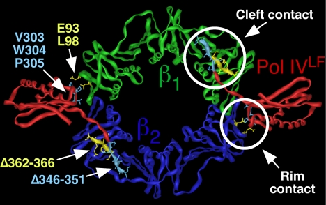
Fig. 1.
Structure of the Pol IVLF-β clamp complex depicting the cleft and rim contacts. The Pol IVLF domain is shown in red, and the clamp is shown in green and blue. Positions of mutations affecting the rim or the cleft contacts between the clamp and Pol IVLF are indicated in yellow and blue, respectively. This figure was generated using imol and PDB coordinates 1UNN (13).

