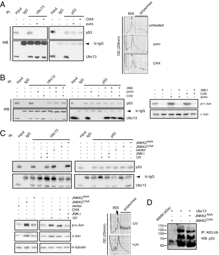
Fig. 2.
Dissociation of Ubc13-p53 complexes is mediated by JNK. (A) U2OS cells were treated with puromycin (puro), cycloheximide (CHX), or incubated with vehicle. Immunoprecipitation reactions were carried out on polysomal fractions using anti-p53, anti-Ubc13, or control isotype-matched IgG antibodies, and further analyzed by Western blot using the indicated antibodies. Right inset represents the UV (254 nm) absorbance profiles of the ribosomal complexes separated on the 10–40% sucrose gradient. 80S indicates the position of monosomes. (B) Left: U2OS cells were treated with puromycin (puro), cycloheximide (CHX), or incubated with a vehicle. Where indicated, the cells were preincubated with a JNK inhibitor (JNK-i). Immunoprecipitations were carried out using anti-p53 and anti-Ubc13 antibodies, or control isotype-matched IgG and the amount of p53 and Ubc13 in the immunoprecipitated material was assessed by Western blot analysis. Inputs were 7.5% of the ribosomal material used in the immunoprecipitation reactions. Right: Levels and phosphorylation of JNK target, c-Jun, upon the indicated treatments were determined by Western blot analysis using c-Jun and anti-phospho-c-Jun (p-c-Jun) antibodies. (C) U2OS cells were untreated, exposed to UV-C light (UV), treated with JNK inhibitor (JNK-i) before the exposure to UV-C light, or transfected with an empty vector (vector), JNKK2CAA or JNKK2AAA constructs. Immunoprecipitations were carried out with anti-p53 and anti-Ubc13 antibodies, or control isotype-matched IgG, and further analyzed by Western blot using the indicated antibodies. Inputs were 10% of the material used for immunoprecipitation. The levels of phosphorylated c-Jun (p-c-Jun), c-Jun, and α-tubulin in the extracts used in the immunoprecipitations reactions were determined by Western blot analysis (lower-left inset). Lower-right inset shows the UV absorbance profiles at 254 nm of the ribosomal gradients from UV treated (+UV) and untreated cells (−UV). (D) U2OS cells were transfected as indicated, and immunoprecipitations from whole cell extracts were carried as described in Methods.

