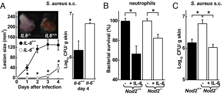
Fig. 6.
Contribution of IL-6 to antibacterial defense. (A) Il6−/− mice (open circles) and WT mice (filled circles) were infected s.c. with 5 × 10⁷ cfu of WT S. aureus and were examined daily for lesion sizes and for bacterial load on day 4. Data are mean (SD); n ≥ 3/group. A representative photograph of the lesions on day 2 is shown in the inset. (B) Freshly prepared bone marrow neutrophils from Nod2−/− mice (open bars) and WT mice (closed bars) were incubated with S. aureus (MOI = 1) with and without IL-6 (50 ng/mL) for 30 min, and surviving bacteria were determined by cfu assay. Data are mean (SE) of 3 independent experiments. (C) Nod2−/− mice (open bar) and WT mice (closed bar) were infected s.c. with 5 × 10⁷ cfu of WT S. aureus. Data are mean (SE); n ≥ 4/group. The indicated group of Nod2−/− mice was treated once daily s.c. with 400 ng of recombinant IL-6. *, P < 0.05.

