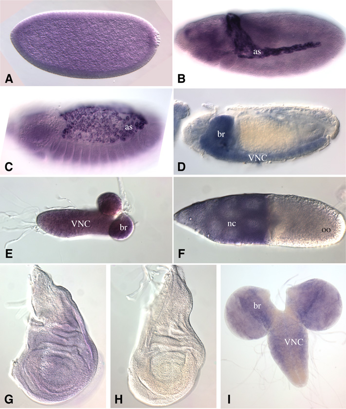
Figure 2. Expression of C1GalTA.
In situ hybridization to Drosophila tissues to detect expression of C1GalTA mRNA. A) Stage 6 embryo. Uniform expression above background is observed in early embryos, which likely includes maternally deposited mRNA. B) Stage 12 embryo. Strong expression is detected in the amnioserosa (as), and weaker expression elsewhere. C) Stage 14 embryo. Strong expression is detected in the amnioserosa. D) Stage 16 embryo. Expression is detected in the VNC and brain (br). E) CNS from first instar larva, expression is detected through out. F) Stage 10 follicle, with strong expression detected in the nurse cells (nc), but not in the oocyte (oo). G) Third instar wing imaginal disc, faint expression is detected. H) Third instar wing imaginal disc hybridized with a sense strand probe as a control, no staining is detected. I) Third instar CNS, C1GaTA is expressed in the brain and VNC.

