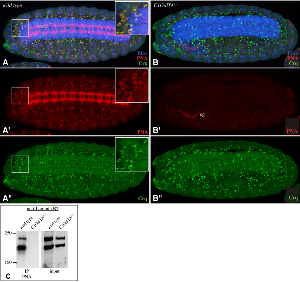
Figure 7. Influence of C1GalTA mutants on PNA staining of hemocytes and Laminin.
A,B show stage 15 embryos stained with PNA (red), anti-Elav (blue), and anti-Crq (green). Panels marked prime show single channels of the embryo above. A) Wild type. PNA staining is detected on hemocytes; inset shows a close-up of the boxed region. B) C1GalTA2.1 mutant; normal PNA staining is lost, although PNA staining is now detected on the salivary gland (sg). C) Western blot using anti-Laminin B2 on lysates from wild-type or C1GalTA2.1 mutant embryos and on material precipitated using PNA-beads. The anti-B2 antibodies cross-react with both Laminin B1 (β, upper band) and Laminin B2 (γ, lower band) (Kumagai et al., 1997).

