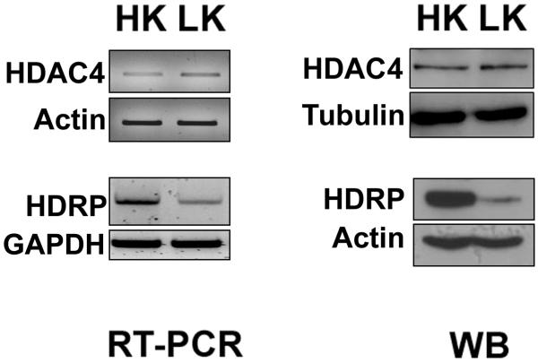
Figure 1. Expression of HDAC4 and HDRP in neurons.
6-7 days old CGNs were treated with HK and LK for 6 hrs. RT-PCR panel; RNA was extracted from cells followed by cDNA synthesis. PCR was performed using primers specific to HDAC4, actin, HDRP and glyceraldehyde-3- phosphate dehydrogenase (GAPDH). Actin and GAPDH were used to demonstrate that similar quantities of cDNA were used. Western blot (WB) panel; cells were lysed and subjected to western blotting using HDAC4 and HDRP antibodies. The same membranes were reprobed with α-tubulin and actin antibodies respectively to demonstrate equal loading.

