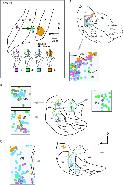
Figure 10.
The locations of electrophysiologically identified injection sites in areas 3a, 3b, 1, and 2 in cortex that was sectioned tangentially (top left box), and coronal sections of the thalamus (A–C) in case no. 4. In the top left box the receptive fields for neurons at the center of each injection site are drawn on illustrations of the hand and color coded to match the area injected. Injections were centered in the representation of distal D3 in areas 3b and 1, and in D2–4 in areas 3a and 2. Patterns of label were similar to those described in case no. 3 (Fig. 8). The injection in area 2 resulted in dense label in VPs, and moderate label in VPl. The injection in area 3a resulted in label in VPl, VPs, and VLp (on rostral sections not shown). In some instances double-labeled cells were observed (grey boxes) in VPl, VPs, and Pla. The distance between sections (A) and (B) is 1400 μm, and between (B) and (C) is 1600 μm. Abbreviations in Table 1. Conventions as in previous figures.

