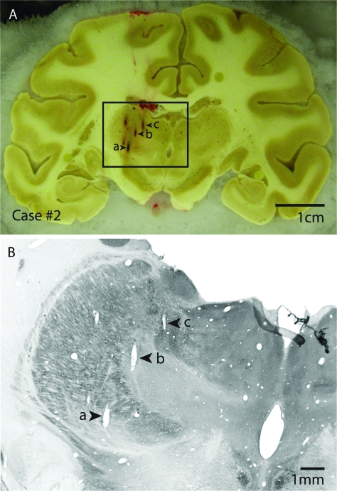
Figure 2.
(A) Block-face image of a coronal section through the cortex and the thalamus in case #2 with electrode penetrations marked a, b, and c. The electrophysiological data reconstruction of this case is shown in detail in Figure 6, and electrode tracks a, b and c correspond to electrode tracks a, b and c between the 2 figures. (B) The section that corresponds to the block-face image in (A) has been processed for CO. Electrode penetrations a, b and c are readily identified in this section, and recording depths can be related to architectonic boundaries of thalamic nuclei. Block-face images combined with histologically processed sections that corresponded to each block-face image were used to guide the reconstruction of electrode penetrations from the dorsal portion of the thalamus through the ventral portion of the thalamus across sections. Dorsal is to the top and lateral is to the left.

