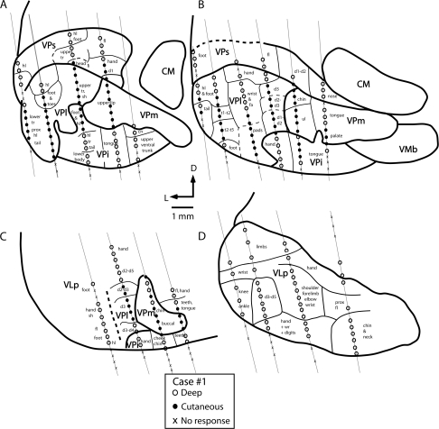
Figure 4.
Coronal reconstructions of electrophysiological recordings from case no. 1. Electrode penetrations and architectonic boundaries have been collapsed across 3 sections for each illustrated section to better appreciate the topographic organization of the somatosensory nuclei of the thalamus. The distance between sections (A) and (B) is 150 μm, (B) and (C) is 350 μm, and (C) and (D) is 150 μm. Several nuclei could be readily distinguished using electrophysiological recording techniques. The ventral posterior nucleus contained neurons that responded to cutaneous stimulation of the contralateral body (VPl) and face (VPm; A–C). Just ventral to VP proper was a second representation of the contralateral body in which neurons responded to stimulation of deep receptors. This nucleus was coextensive with the architectonically defined VPi. The third nucleus is VPs (A and B). Neurons in VPs responded to stimulation of deep receptors of the contralateral body. There was a gross topographic organization of VPs that mirrored that of VPl and VPm. Finally, at the rostral boundary of VP proper, neurons responded to stimulation of deep receptors of the contralateral body; this representation was coextensive with the architectonically defined VLp (D). Thick lines denote architectonic boundaries of each nucleus and thin lines separate different body part representations within each nucleus. Dashed lines mark approximate architectonic boundaries. This series of sections and series on all remaining figures are drawn caudal (A) through rostral (D). See Table 1 for abbreviations. Conventions as in previous figures.

