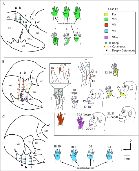
Figure 6.
Recording site progressions for neurons through coronal sections of the thalamus in case #2 (A–C) in tracks a, b and c, and corresponding receptive fields for neurons at those sites (body part diagrams on the right). As in the previous case (Fig. 5) neurons in VPs (A) responded to stimulation of deep receptors and receptive fields on the hand (green) were large. As recording sites progressed ventrally, in VPl (orange) and VPm (pink; B) neurons responded to stimulation of cutaneous receptors on the contralateral hand and face, and receptive fields were much smaller. One electrode track (b) was at the border of VPm and the cell-sparse, fingerlike protrusion of VPi, and receptive fields at a number of sites were split (e.g., r.f. 18, 20, 21). Receptive fields on the face were small and neurons responded to cutaneous stimulation (in VPm), and receptive fields on the hand were large and neurons responded to stimulation of deep receptors (in finger-like extensions of VPi). The recording sites located in the most ventral portion of the dorsal thalamus, in VPi (blue; C), contained neurons that responded to stimulation of deep receptors and receptive fields for these neurons were relatively large and encompassed all or most of the hand (see blue r.f in C). In electrode track c, some recording sites were in Pla (yellow). Neurons here responded to stimulation of cutaneous receptors on the contralateral body, and receptive fields were on the face, hand, or both the face and the hand. Sections of the thalamus drawn on the left are composites of 4 sections each, and each composite covers 800 μm. See Table 1 for abbreviations. Conventions as in previous figures.

