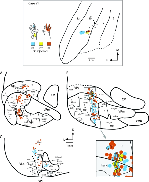
Figure 8.
The location of electrophysiologically identified injections sites in area 3b in cortex that was sectioned tangentially (top box), and coronal sections of the thalamus (A–C) in case #1. In the top box, the receptive fields for neurons at the center of each injection site are drawn on illustrations of the hand and color coded to match the tracer injected. All injection sites are restricted to portions of the hand representation. The injection of FB was centered in the representation of glabrous middle D3, the injection of DY was centered in the representation of the proximal glabrous thenar pad, and the injection of FR was centered in the representation of the dorsal digits 3–5. Labeled cells resulting from these injections are depicted below and their direct relationship to electrophysiologically defined locations in the thalamus is depicted in sections A–C. These sections correspond to similarly labeled sections in Figure 4. These data indicate that a large amount of convergence is observed in thalamic projections to area 3b in that a given body part representation in area 3b, such as the middle glabrous D3, receives input not only from the same body part representation in VPl, but from other representations in VPl such as digit 2 and portions of the hand. Further, a few labeled cells were observed in the chin representation of VPm. Convergence is also observed across nuclei in that 3 separate nuclei (VPl, VPs, and VPi) project to a single location in area 3b. Finally, divergence is seen at the cellular level in that in some instances the same cells projected to 2 separate representations in area 3b. The large shaded box at the lower right of this figure is an enlarged image of the shaded square shown in B. Double- and triple-labeled cells in VPs and VPl are indicated by multicolored dots. See Table 1 for abbreviations. Conventions as in previous figures.

