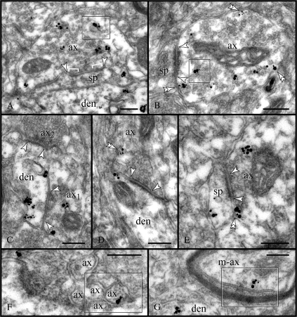
Figure 7.
Presynaptic expression of RGS4. (A, B) Labeling was predominantly found in the axoplasm of non-Glut–like varicosities; note the association with endoplasmic vesicles (frames). Relatively few particles labeled the axolemma (double arrowheads), which may indicate a high turnover rate for RGS4 or a corresponding GPCR. Synapses (between arrowheads) were mainly onto immunonegative spines. (C) Convergent synapses (arrowheads) of non-Glut–like and Glut-like axons (ax1 and ax2, respectively) onto a labeled dendrite. Note the atypical particle distribution in the dendrite as immunoreactivity is not associated with PSDs (see Fig. 5). (D, E) Glut-like axons establishing axodendritic and axospinous synapses (arrowheads) displayed low expression of RGS4 and minimal axoplasmic labeling. Rather, RGS4 was associated with extrasynaptic and perisynaptic membranes (double arrowheads). Nonmyelinated (F) and myelinated (G) axon intervaricose segments were also labeled (frames). ax; axon; den, dendrite; m-ax, myelinated axon; and sp, spine. Scale bars: 200 nm.

