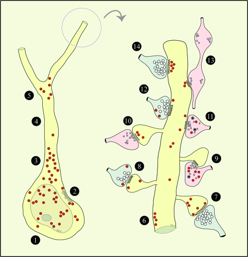
Figure 9.
Schematic depiction of RGS4 expression patterns in the perisomatic region and in a spinous dendrite. For brevity, all axodendritic synapses are shown targeting a pyramidal dendrite, although they could potentially engage nonpyramidal dendrites as well. Axons with Glut-like features are shown in blue; non-Glut–like axons are in pink. Red polygons depict RGS4 labeling. 1) Soma—low and high RGS4 in cytoplasm and nucleoplasm, respectively. 2) Soma—RGS4 subjacent to SSC-lined membranes. 3) Proximal apical dendrite—very high RGS4 in cytoplasm. 4) Distal apical dendrite—paucity of immunoreactivity*. 5) First- to second-order dendrites—RGS4 in bifurcation point. 6) Nonsynaptic membranes/axon appositions—RGS4 subjacent to plasmalemma. 7) Asymmetric axospinous synapse—low RGS4 in spine subjacent to PSD, high RGS4 perisynaptically and extrasynaptically. 8) Asymmetric axospinous synapse—low RGS4 in spine extrasynaptically; low RGS4 in axon perisynaptically and extrasynaptically. 9) Symmetric axospinous synapse—diffuse high RGS4 in axon. 10) Symmetric axospinous synapse—RGS4 in spine subjacent to PSD. 11) Symmetric axodendritic synapse—low RGS4 in dendrite extrasynaptically; diffuse low RGS4 in axon. 12) Asymmetric axodendritic synapse—low RGS4 in dendrite extrasynaptically; low RGS4 in axon extrasynaptically. 13) En passant symmetric axodendritic synapse—RGS4 in dendrite subjacent to PSD. 14) Asymmetric axodendritic synapse—very high RGS4 in dendrite subjacent to PSD. *Not in all cells.

