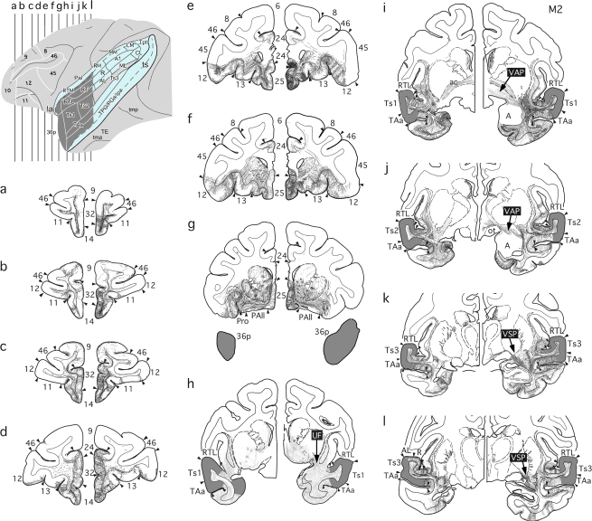
Figure 4.
Lateral surface view and coronal sections (a–l) illustrating anterograde tracer injections (dark gray) in rSTG, with “opened” lateral and superior temporal sulci shown in blue, and resulting fiber and terminal label in case M2. In the intact right hemisphere, labeled efferent fibers from the injected STG region joined 3 major pathways: One coursed through the uncinate fasciculus (h) and led to dense terminal label in the ventral medial frontal cortex (a-g), whereas a second and third followed the ventral amygdalofugal (i, j) and ventral striatum pathways (k, l), leading to dense terminal label in the medial thalamus. In the hemisphere with the MTL removal, all 3 efferent pathways were interrupted resulting in substantially less dense label in these 2 target areas. A, amygdala; 13, 14, 24, 25, 32 Brodman's cytoarchitectonic areas; ai, inferior arcuate sulcus; as, superior arcuate sulcus; p, principal sulcus; PAll, frontal periallocortical area; Pro, proisocortex; UF, uncinate fasciculus; VAP, ventral amygdalofugal pathway; VSP, ventral striatum pathway; see Figure 1 for abbreviations.

