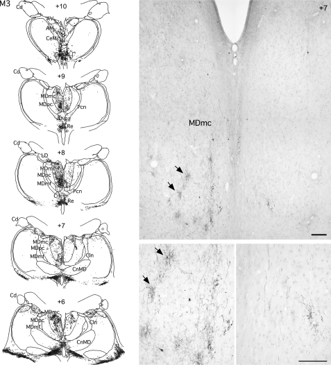
Figure 5.
Coronal sections and photomicrographs from case M3 showing the distribution of anterograde label in the medial thalamus after bilateral BDA injections in the rSTG. Photomicrographs at 2 different magnifications illustrate the patchy distribution of the label in the magnocellular portion of the medial dorsal nucleus of the thalamus. Note the decreased density of label in the right hemisphere with the MTL ablation. Scale bar: 250 μm. Cln, central lateral thalamic nucleus; CnMD, centrum medianum thalamic nucleus; Cd, caudate nucleus; Pcn, paracentral nucleus; CeM, central medial thalamic nucleus; AD, anterior dorsal thalamic nucleus; AM, anterior medial thalamic nucleus; AV, anterior ventral thalamic nucleus; Cif, central inferior thalamic nucleus; Cim, central intermedial thalamic nucleus; MDmc, medial dorsal thalamic nucleus magnocellular division; MDmf; medial dorsal thalamic nucleus multiformis division; MDpc, medial dorsal thalamic nucleus parvocellular division; Pcn, paracentral nucleus; Pf, parafascicular nucleus; Re, Reuniens; Ro, Rotundus; sf, subfascicular nucleus.

