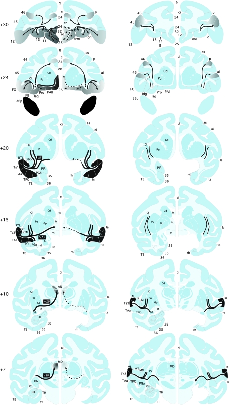
Figure 9.
Summary diagrams of the major fiber pathways examined in this study. Left column of coronal sections illustrates projections from the rSTG to the frontal cortex and medial thalamus. The series of sections depicts each pathway's trajectory, from its origin in the injected cortical tissue (shown in black), to the course it follows through the white matter (black lines of varying thickness, representing graded size of projections), to its destination in cortex or thalamus (shown in shades of gray, representing graded density of terminal label). The projection from rSTG to the frontal cortex travels through the uncinate fasciculus (UF, +20). A major branch of this pathway continues medially to course below the striatum on the unoperated side (left hemisphere, solid line with arrowhead at +20) and then turns rostrally to terminate in medial and orbital frontal cortical areas on this side (left hemisphere, solid lines and dark gray shading at +24 and +30), with the highest density of terminal label in the ventral medial frontal cortical areas. Aspiration of the MTL (right MTL lesion at +7 through +20) transected this medial branch of the UF (right hemisphere, dashed line with arrowhead at +20) and, as a result, there was only sparse terminal label in the ventral medial frontal cortical areas on this side (right hemisphere, dashed lines with arrowheads and light gray shading at +24 and +30). Some fibers comprising the UF do not continue medially from their origin but, instead, turn dorsally and rostrally to travel through the external and extreme capsules (left hemisphere, solid lines at +15 and +20), after which they converge with the more medial branch of the uncinate fasciculis to terminate in mid and lateral orbital areas as well as in small frontal areas dorsally (left hemisphere, thin solid lines with arrowheads and light gray shading at +24 and +30). These branches of the UF escaped damage on the lesioned side and so the terminal label at their destinations was unaffected. The MTL removal also transected the caudally projecting fibers that join the VAP (at +15) and VSP (at +10 and +7), resulting in reduced terminal label in the medial thalamus (AN and MD at +10 and +7). Right column of coronal sections depicts projections to the frontal cortex from the injected areas of the auditory belt and parabelt divisions of the caudal STG. The MTL removal had no effect on these fiber pathways (solid black lines in all coronal sections, and so the terminal label was also unaffected (equivalent gray shading in both hemispheres at +24 and +30). AN, anterior thalamic nuclei; ai, arcuate sulcus, inferior; as, arcuate sulcus, superior; cc, corpus callosum; ci, cingulate sulcus; Cl, claustrum; Iag, insula, agranular subdivision; Idg, insula, dysgranular subdivision; los, lateral orbital sulcus; orl, lateral orbital sulcus; orm, medial orbital sulcus; FO, frontal operculum; fx, fornix; Gp, globus pallidus; H, hippocampus; Pu, putamen; sf, subfascicular nucleus; MD, medial dorsal thalamic nucleus; see Figure 1 for abbreviations.

