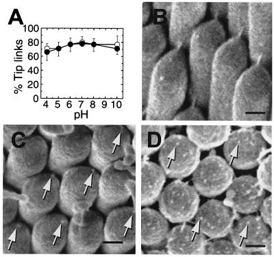
Figure 5.
Response of tip links to chemical treatment. (A) Chicken basilar papillae were exposed to solutions of varying pH and either 4 mM (●) or 1.4 μM (○) CaCl2. For each point, tip links from 14–16 hair bundles from 3–4 papillae were counted from SEM images; each point corresponds to ≈1,000 tip-link positions. Means ± standard error are plotted. Bundles were chosen from all regions of the papilla. (B–D) Field-emission SEM images of chicken basilar-papilla hair bundles before and after BAPTA treatment. (B) Control. (C) Immediately after BAPTA treatment; remnants are seen where tip links formerly anchored (arrows). (D) 2 h after BAPTA treatment, during tip-link regeneration. Protuberances on the surface of each stereociliary tip are indicated by arrows. [Scale bars: B = 200 nm (applies to C and D).]

