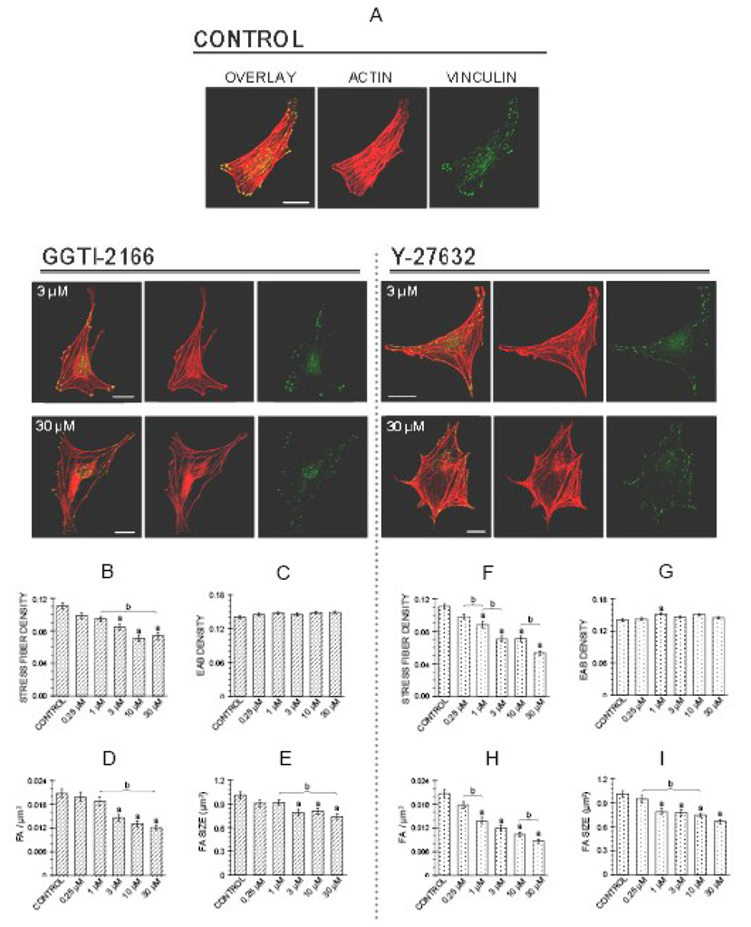
Figure 2.
Geranylgeranyl transferase I inhibitor GGTI-2166 (left panel) or Rho kinase inhibitor Y-27632 (right panel) decrease stress fiber density, focal adhesion density and focal adhesion area in MC3T3-E1 cells. Treatments were for 6 hr in BSA medium. A: Representative confocal micrographs of cells showing actin stress fibers and focal adhesions. Bars represent 25 µm. B–I: Quantification of parameters. Values are means +/− SEM of responses measured in 3 independent experiments in which 40 cells were imaged and quantified per group. There are no statistically significant differences in edge actin bundle (2C, 2G), cellular area, or circularity (data not shown). Different letters indicate statistically significant differences (95% Tukey confidence interval) from control (a) and between groups (b).

