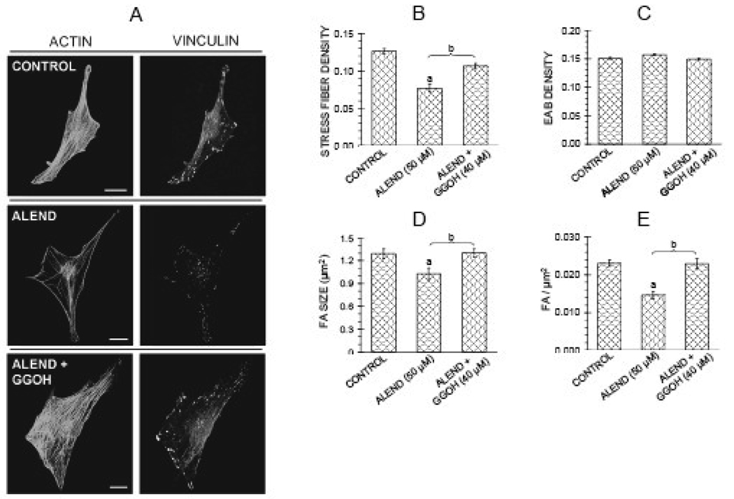
Figure 3.
Alendronate (50 µM) decreases stress fiber density, focal adhesion density, and focal adhesion area in MC3T3-E1 cells, and effects are antagonized by geranylgeranyl group donor geranylgeraniol (GGOH) (40 µM). Treatments were for 22 hr in FBS medium. A: Confocal micrographs of cells showing actin stress fibers and focal adhesions. Bars represent 25 µm. B–E: Quantification of parameters. Values are means +/− SEM of responses measured in 2 independent experiments in which 40 cells were imaged and quantified per group. Different letters indicate statistically significant differences (95% Tukey confidence interval) from control (a) and between groups (b). There are no statistically significant differences in edge actin bundle (3C), cellular area or circularity (data not shown).

