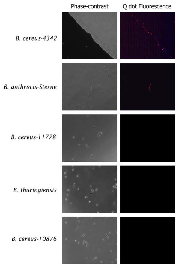
Figure 5.
Microscopy-based sensitivity analysis of PlyG-P3 peptide binding. Bacterial cultures of Bacillus cereus 4342, B. anthracis-Sterne, B. cereus-11778, B. thuringiensis, and B. cereus-10876 were incubated with PlyG-P3 and the complex was detected by incubating with strepatividin-conjugated quantum dots. Peptide binding was detected by the fluorescing Q dots using appropriate UV filters (605 nm) under a Nikon microscope, either in phase-contrast (panels on left) or fluorescence (panels on right) fields. Note that even a single bacterium is visible in the B. cereus-Sterne field.

